Hamamatsu Photonics K.K. heeft een nieuwe 3D-fluorescentiescanmethode ontwikkeld, “Zyncscan™” genaamd, die een X-Z-vlakscan uitvoert met een lichtfolie voor celgebaseerde fluorescentietests in microplaten
HAMAMATSU, Japan-(BUSINESS WIRE)-Hamamatsu Photonics K.K. (TOKYO:6965) heeft een nieuwe 3D-fluorescentiescanneringmethode ontwikkeld, de zogenaamde “Zyncscan™”, die x-z-vlakke scans uitvoert met behulp van een lichtblad voor celgebaseerde fluorescentie-analyses in microplaten. Deze technologie biedt een x-z vliegtuigscan met behulp van een light-sheet voor een hele 96/384/1536-well microplaat, zodat gebruikers 3D-fluorescentie beelden van hele wells in xy te verkrijgen: 2-3 urn en z: 6-7 urn voxel resolutie voor ≤ 300 urn dikte van de putbodem binnen een paar minuten per kleur. Deze technologie maakt het ook mogelijk voor ultra-hoog niveau scheiding van celfluorescentiesignalen van de achtergrond, die het mogelijk maakt om fluorescentiecel beelden te verkrijgen in de celkweekmediums met serum en met fluorescerende kleurstoffen (dat wil zeggen, geen noodzaak om uit te wassen fluorescerende kleurstoffen).
Hamamatsu Photonics K.K. Has Developed a Novel 3D Fluorescence Scanning Method Called “Zyncscan™“ That Performs an X-Z Plane Scan With a Light-Sheet for Cell-Based Fluorescence Assays in Microplates
HAMAMATSU, Japan–(BUSINESS WIRE)– Hamamatsu Photonics K.K. (TOKYO:6965) has developed a novel 3D fluorescence scanning method called “Zyncscan™” that performs x-z plane scans using a light-sheet for cell-based fluorescence assays in microplates. This technology provides an x-z plane scan using a light-sheet for a whole 96/384/1536-well microplate, allowing users to obtain 3D fluorescence images of whole wells in xy: 2-3 μm and z: 6-7 μm voxel resolution for ≤ 300 μm thickness from the well bottom within a few minutes per color. This technology also allows for ultra-high level separation of cell fluorescence signals from the background, which makes it possible to obtain fluorescence cell images in the cell culture mediums containing serum and with fluorescent dyes (i.e., no need to wash out fluorescent dyes).
This press release features multimedia. View the full release here: https://www.businesswire.com/news/home/20190609005009/en/

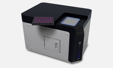
CYTOQUBE(TM) (Light-Sheet Microplate Cytometer) Prototype equipped with Zyncscan(TM) technology (Photo: Business Wire)
Background of development
In drug discovery and development, there is a rapidly growing need to use more physiologically-relevant in vitro cell culture models. These include primary cells, patient-derived cells, human iPSC-derived disease-modeling cells, co-cultures of different types of cells, and 3D cultures such as spheroids and organoids. To develop and advance phenotypic assays and screenings using these physiologically-relevant cellular models, new types of instruments needed to be developed to facilitate high-throughput fluorescence imaging and measurement of heterogeneous 2D and 3D cell cultures. To meet this need, we have developed ZyncscanTM technology that performs an x-z plane scan utilizing a light-sheet for a microplate.
Features of the cell-based fluorescence assays in microplate using Zyncscan™ technology
| 1) | High-speed fluorescence cytometry for 2D monolayer heterogeneous cell culture | |
| A single 120-second scan (one color) for a whole 96/384/1536-well microplate enables fluorescence cytometry of all individual cells in all wells. | ||
| 2) | Single spheroid analyses using depth information (diameter of spheroid; ≤ 200 μm) | |
| A single scan (in just a few minutes) for a microplate enables fluorescence images of spheroids (single or multiple spheroids in a well) in all wells in a 96/384/1536-well microplate. From the images, the entire fluorescence intensity, thickness and volume of each individual spheroid are estimated. | ||
| 3) | Fluorescence images in 3D view (≤ 300 μm) | |
| With a single scan (a few minutes) obtain 3D fluorescence images (300 μm from the bottom of a well) of all wells in a plate. The optical resolution of the image is comparable with the 2x objective of a conventional fluorescence microscope in the x- and y-axes and a 10x objective of confocal fluorescence microscope in the z-axis. | ||
| 4) | In-medium / No dye washout measurement | |
| Measure cellular fluorescence in the medium containing serum and with fluorescent dyes (no need to wash out fluorescent dyes), allowing cells to remain healthy throughout the experiment. | ||
Future outlook
Hamamatsu Photonics K.K. has been developing a fluorescence instrument for cell-based assays equipped with Zyncscan™ technology, CYTOQUBE™, a Light-Sheet Microplate Cytometer. We plan to initiate validation and application experiments in several research sites in drug discovery and development by early 2020. CYTOQUBE™ is planned to be released by the end of the next fiscal year (September 2020).
We will showcase a prototype of the CYTOQUBE™ (Light-Sheet Microplate Cytometer) at the following conferences.
| WPC (World Preclinical Congress) |
| June 17-20, 2019, Boston, MA, USA |
| CYTO |
| June 22-26, 2019, Vancouver, BC, Canada |
Please visit here for more detailed information.
https://www.hamamatsu.com/all/en/news/featured-products_technologies/2019/20190610000000.html
Hamamatsu Photonics K.K.
Hamamatsu Photonics K.K. is a leading manufacturer of photonics devices. We design, manufacture, and sell optical sensors, light sources, optical components, cameras, photometry systems, and measurement/analysis systems.
Web site
View source version on businesswire.com: https://www.businesswire.com/news/home/20190609005009/en/
Contacts
Technical Contacts details
Hamamatsu Photonics K.K, System Division
Business Promotion Department
812, Joko-cho, Higashi-ku, Hamamatsu City, 431-3196 Japan
email: export@sys.hpk.co.jp

